Work Packages
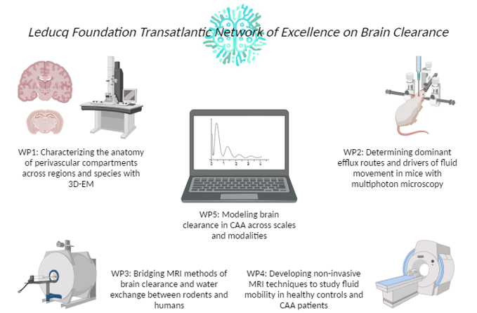
WP 1: Characterizing the anatomy of perivascular compartments across regions and species.
This work package (WP) will leverage existing electron microscopy (EM) datasets in rodents to provide the first comprehensive characterization of the perivascular compartments along the cerebrovascular tree. It will also generate novel 3D-EM data in freshly resected gene-edited CAA rodents and human brain tissue needed to reliably map the ultrastructural anatomy of vascular cells, basement membranes, astrocytic endfeet, and perivascular spaces (PVS) in normal appearing and CAA-affected vessels. Assessing vessels at the time when CAA starts to develop will provide important clues on the anatomical pathways of physiological Aβ clearance.
WP 2: Determining dominant efflux routes and drivers of fluid movement in mice.
This WP will use minimally invasive 2-photon microscopy in wild-type and novel gene-edited CAA mice to define physiological clearance routes, rates, and drivers. We will use uncageable fluorescent dyes and Aβ to define mobility in vivo after achieving steady-state tracer concentrations through chronic, slow infusion via the intracerebral ventricles. This minimizes introduction of intracranial pressure increases or artificial diffusion gradients present with bolus tracer injections. Further, we will use transgenic labeling strategies to measure PVS dimensions without exogenous dyes and to clarify physiologic driving forces that influence tracer clearance and PVS dynamics.
WP 3: Bridging MRI methods of Brain Clearance and water exchange between rodents and humans.
This WP will adapt existing contrast agent (CA)-based MRI techniques in the healthy rat and a recently developed novel gene-edited CAA rat model, to define macroscopic efflux routes from the brain. Furthermore, by adapting human, non-invasive MRI sequences that capture water exchange dynamics for rodent imaging and validating these measures against gold-standard CA-based imaging, this WP will build the translational bridge between rodent and human studies. A key knowledge gap is how CAA affects global Interstitial Spinal Fluid (ISF) efflux from the parenchyma to the Cerebrospinal Fluid (CSF), and the clearance pathway to extracranial lymph nodes.
WP 4: Developing non-invasive MRI techniques to study fluid mobility in healthy controls and CAA patients.
This WP focuses on the optimization and application of recently developed non-invasive MRI sequences to capture CSF/ISF mobility and water exchange dynamics as a surrogate for brain clearance in the human brain. These innovative methods will define, for the first time at the level of the PVS, fluid mobility in healthy volunteers, then evaluate changes in patients with sporadic CAA and individuals with hereditary (Dutch-type) CAA. We will furthermore test whether manipulating the relevant driving forces established in rodents can be translated to humans, thus establishing key proof-of-concept data for future clinical trials.
WP 5: Modeling Brain Clearance in CAA across scales.
This WP will develop new computational models of brain clearance by integrating experimental findings from WP 1-4. Based on new anatomical and transport data across physiological states, these models will permit the prediction of physiological and pathological modifiers of perivascular brain clearance across a wide range of conditions, also those not accessible through experimental manipulations. An important knowledge gap is the dynamics of clearance at the mesoscopic scale and in grey vs. white matter.