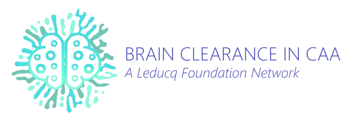
Amyloid-β stained blood vessel in a post-mortem human brain section.
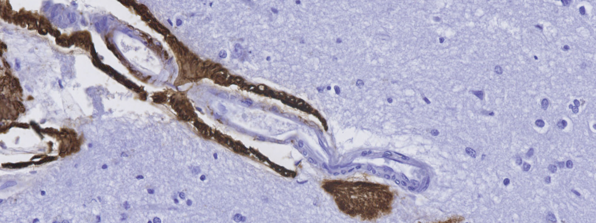
Blood vessel with CAA in a mouse, imaged with in vivo two-photon microscopy.
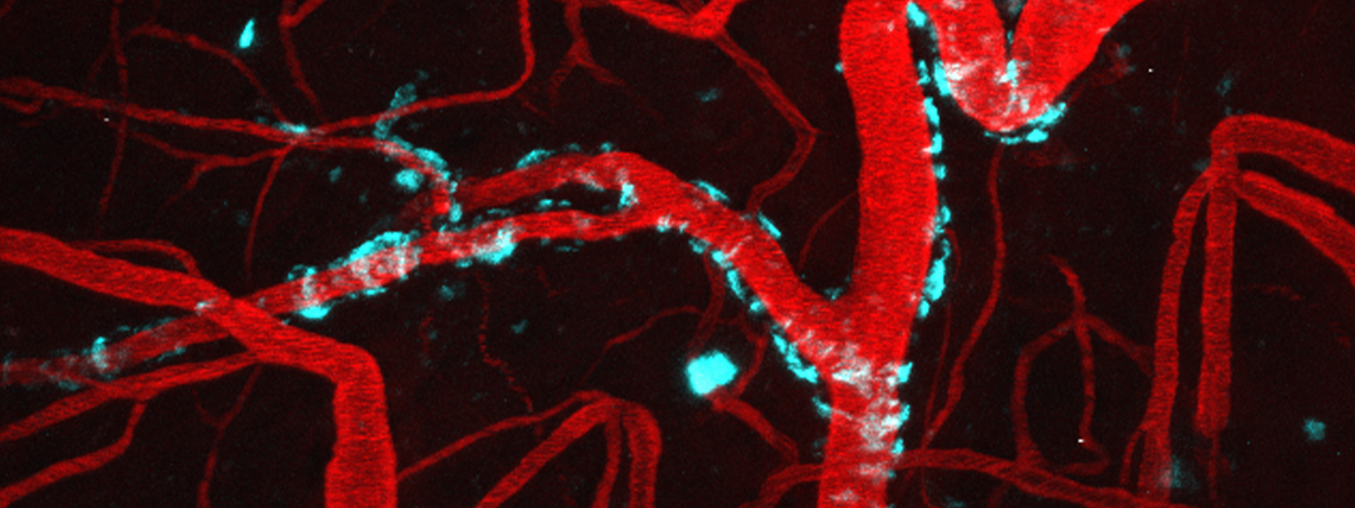
Immunofluorescence of a blood vessel with reactive astrogliosis.
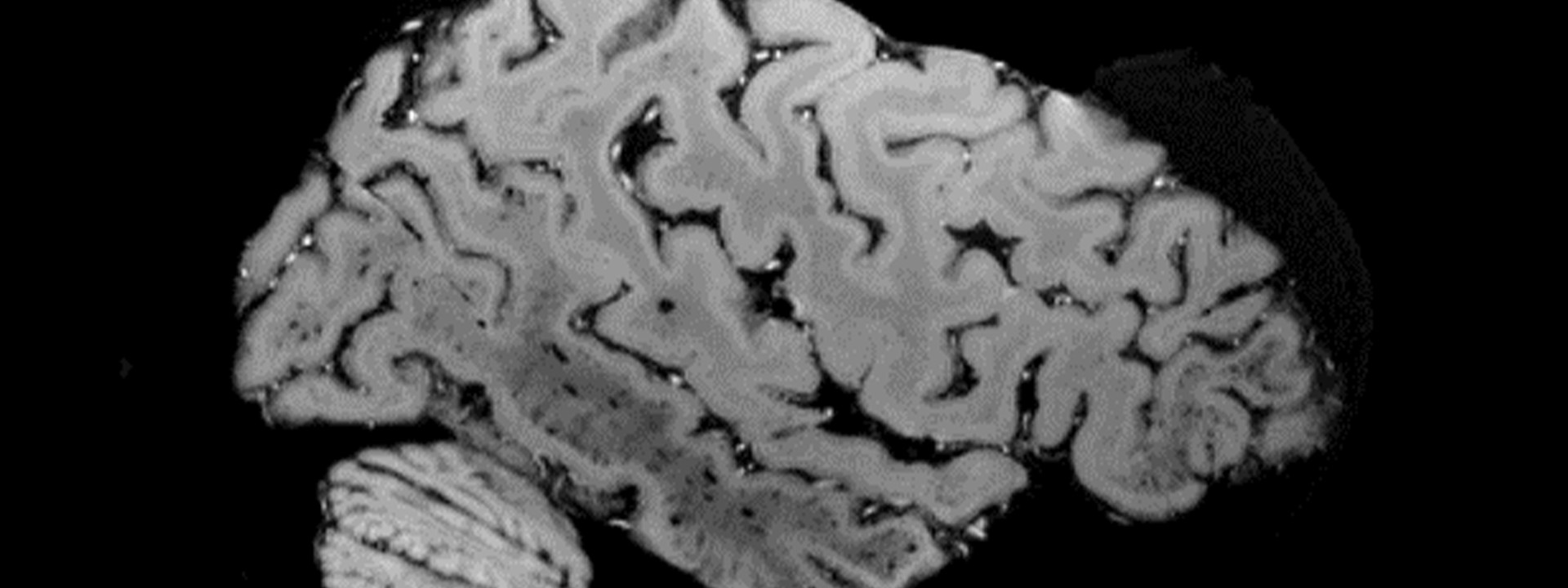
Ex vivo 3T MRI of a formalin-fixed hemisphere of a patient with CAA.
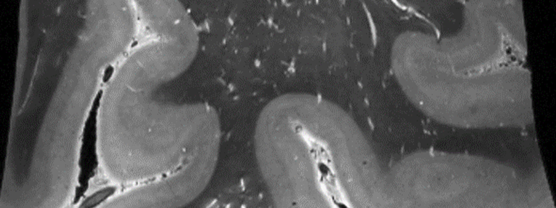
Ex vivo 7T MRI of a brain sample with enlarged perivascular spaces.
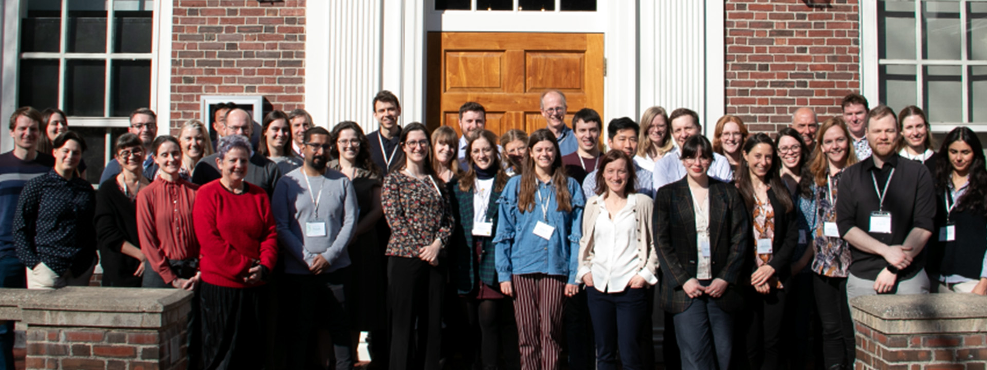
Leducq members at the Kick-off meeting in Boston at the Harvard Faculty Club.
Welcome to the Leducq Foundation Transatlantic Network of Excellence on Brain Clearance!
This Network brings together internationally recognized leaders in the fields of Cerebral Amyloid Angiopathy (CAA) and brain clearance, who sometimes have opposing views on the underlying physiological principles of brain clearance, with experts using innovative imaging techniques to measure and manipulate brain clearance in animal models and humans. Together we aim to elucidate fundamental unanswered questions related to brain clearance by focusing on both healthy brains and brains affected by CAA. This is done in a translational (from rodent to man), multimodal (from electron and multi-photon microscopy to MRI), and multidisciplinary (from mechanistic modeling to clinical) fashion.